Unique microscopic images provide new insights into ionic liquids

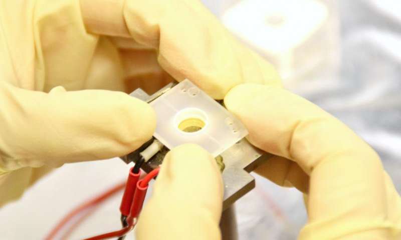
The researchers use a special sample holder to investigate the ionic liquids under the microscope. Credit: CAU, Denis Schimmelpfennig
To directly observe chemical processes in unusual, new materials is a scientific dream, made possible by modern microscopy methods: researchers at Kiel University have, for the first time, captured video images of the attachment of molecules in an ionic liquid onto a submerged electrode. The images from the nanoscale world provide detailed information on the way in which chemical components reorganise when a voltage is applied. New findings based on this information may lead to improved batteries and more energy efficient coating technology or solar engineering.
Ionic liquids are organic salt melts, which may even be fluid at room temperature, although they contain no water. It is exactly this point that makes them so interesting for numerous experiments and industrial processes. This is because water is electrolytically dissociated at electrodes even at small voltages, which blankets and hinders other, technically important, electrochemical reactions. In addition, the water molecules encase the ions and interfere in numerous chemical processes. In ionic liquids, which consist of ions only, completely new reactions are therefore possible.
Ionic liquids have been a hot field of research in recent years, leading to the discovery of a whole range of new compounds. Their technological applications are manifold: As electrolytes in batteries, fuel cells or dye solar cells and as a galvanic bath for the deposition of thin aluminium coatings or semi-conductor materials. The fact that they operate at room temperature makes them easier to handle for numerous applications and saves energy on top.
However, at present almost no data are available on how electrochemical reactions in ionic liquids operate at the molecular level or how the molecules are arranged on the surface of the electrode. While in aqueous liquids this has been studied for decades by modern microscopy methods, similar studies in ionic liquids have been largely unsuccessful: "The molecules often simply move too fast for conventional instruments", says Professor Olaf Magnussen of Kiel University. Using a self-built scanning tunnelling microscope his team was now able to track down this mystery.
Microscopic video images of a negatively charged gold electrode in an ionic liquid. The fluctuating square pattern is formed by the liquid's BMP molecules, which attach onto the metallic surface in an ordered arrangement under these conditions. Credit: AG Magnussen
Video sequences recorded by Magnussen's co-worker Dr Rui Wen reveal how the liquid's molecules, less than a nanometre in size, react when a voltage is applied to a gold electrode. If the surface is uncharged, the molecules display a response typical for liquids: they are disordered and highly mobile. As the voltage increases the molecules lay down flat on the surface and form rows, before they finally reorient to an erect arrangement. At the same time, they become less and less mobile. "The images are unique and help us to develop theories to better describe the electrode processes in ionic liquids", says physicist Magnussen. "This is important not only for basic research, but also for concrete applications."
Unique microscopic images provide new insights into ionic liquids

The researchers use a special sample holder to investigate the ionic liquids under the microscope. Credit: CAU, Denis Schimmelpfennig Under the leadership of Professor Olaf Magnussen, Wen's research on ionic liquids took place on the group's home-build video scanning tunnelling microscope. Credit: CAU, Denis Schimmelpfennig
Read more at: http://phys.org/news/2015-04-unique-microscopic-images-insights-ionic.html#jCp
